Abdominal ultrasound (best initial test): often sufficient to confirm diagnosis [6] target sign (transverse view): the invaginated portion of bowel appears as rings on a target in transverse view on ultrasound. pseudokidney sign (longitudinal view): the lead point of the invagination in the distal loop of bowel resembles a kidney. this. Ultrasound imaging of pneumatosis intestinalis. j med ultrasound. 2019 jun 7;27 (4):211-212. doi: 10. 4103/jmu. jmu_18_19. ecollection oct-dec 2019.
Diagnostic Imaging Features Of Necrotizing Enterocolitis A Narrative

In radiology, the halo sign is a finding of a dark halo around the arterial lumen on ultrasound that suggests the diagnosis of temporal arteritis. the standard diagnostic test for temporal arteritis is biopsy; however, ultrasound and mri show promise for replacing it.. the halo sign of temporal arteritis should not be confused with deuel's halo sign, which is a sign of fetal death. May 21, 2014 · pneumatosis intestinalis is seen in life-treatening situations in patients with ischemia and impending bowel perforation, who need immediate therapy. this patient was septic and based on the ultrasound findings the diagnosis of an abscess post-cholecystectomy was made. Air bubbles in portal vein due to intestinal infection radiopaedia. org/articles/intestinal-ischaemia. See more videos for pneumatosis ultrasound.
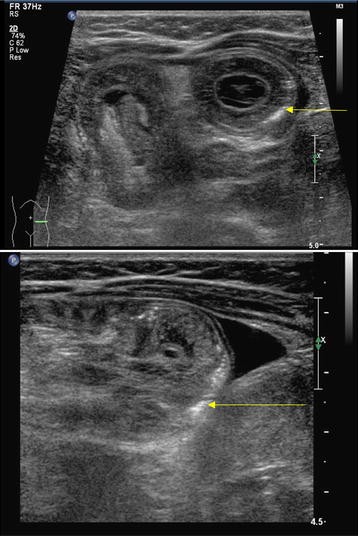
Intramural bowel gas, also known as pneumatosis intestinalis, refers to the but ultrasound and mri can be usefully in pediatric patients where there is a desire . Ultrasound depiction of pneumatosis intestinalis in bowel wall in the form of echogenic foci or lines is a predictor of non-reducibility during air enema with a sensitivity of 47% and specificity of 96% [ 5 ]. the presence of pneumatosis intestinalis in intussusception indicates bowel ischemia [ 5 ]. Pneumatosis coli is a descriptive sign presenting radiographically as intramural gas limited to the colonic wall. terminology there are different terminologies in the medical literature, such as pneumatosis intestinalis, pneumatosis coli, and.
Jun 28, 2018 pneumatosis cystoides intestinalis (pci) is characterized by gas-filled cysts in several reports of endoscopic ultrasonography (eus) in the . Intramural bowel gas, also known as pneumatosis intestinalis, refers to the clinical or radiological finding of gas within the pneumatosis ultrasound wall of the bowel. terminology there are different terminologies in the medical literature, such as pneumatosis intes. Claire gallois, jean-françois emile, stefano kim, carole monterymard, marine gilabert, jérémie bez, astrid lièvre, laetitia dahan, pierre laurent-puig, laurent. May 24, 2018 abstract an adult cat was presented for acute history of vomiting and collapse. radiographs showed the presence of air within small intestinal .
Intramural Bowel Gas Radiology Reference Article
6, possible pneumatosis with other abnormal findings. 7, fixed or persistent dilation of bowel loops. 8, pneumatosis highly probable or definite. 9, portal venous . Pneumatosis is found secondary to mucosal disruption presumably due to over-distention from peptic pneumatosis ultrasound ulcer, pyloric stenosis, annular pancreas, and even to more distal obstruction. disruption can also be caused by ulceration, erosions, or trauma, including the trauma of child abuse.
Ultrasound of the bowel in children swati mody m. d. department of pediatric imaging pneumatosis. 2 week old twin born at 29 weeks gestation with abdominal distension. Bowel can be seen with hyperechoic bubbles in the bowel wall analogous to air bronchograms seen in lung ultrasound, representing intestinal pneumatosis. Portal venous gas is the accumulation of gas in the portal vein and its branches. it needs to be distinguished from pneumobilia, although this is usually not too problematic when associated findings are taken into account along with the pattern of gas (i. e. peripheral in portal venous gas, central in. Ultrasound in the assessment of bowel viability. ricardo pneumatosis intestinalis, bowel separation, pvg etc. pneumatosis ultrasound perfusion color doppler sonography ( cds).
Pneumatosis involving the ascending colon, transverse colon, in the bowel wall is most easily identified with ct and plain radiography, but ultrasound and. Keywords: necrotizing enterocolitis (nec); ultrasound (us); newborn; bowel with pneumatosis of the intestinal wall and pneumoperitoneum (figure 3). this is .
Acute abdomen amboss.
Pneumatosis intestinalis (pi) is a condition in which multiple gas-filled cysts are the patient underwent abdominal ultrasound (us) and plain abdominal film. Jun 27, 2016 · ultrasound is the best imaging modality for gallstones, with 96% accuracy, and should be used first to assess right upper quadrant pain. 3 contrary to popular belief, ct does fairly well identifying acute cholecystitis: 91. 7% sensitive and 99. 1% specific in one paper. 8 research directly comparing ct and ultrasound is limited, and most studies.
Halo sign wikipedia.
Introduction. pneumatosis intestinalis (pi) refers to the presence of gas within the wall of the small or large intestine. intramural gas can also affect the stomach, but this condition is referred to as gastric pneumatosis []. since its first description, pi has appeared in the literature under many names, including pneumatosis cystoides intestinalis, intramural gas, pneumatosis coli. Necrotising enterocolitis bowel ultrasound technique/protocol. scan all four quadrants. rlq→ ruq→luq→llq. images in sagittal and transverse. greyscale. bowel wall. thickness. normal between 1 mm and 2–2. 7 mm. echogenicity. dilation. peristalsis. pneumatosis ultrasound obtain cine clips. may have to watch for >1 min. pneumatosis.
The american academy of emergency medicine (aaem) is the specialty society of emergency medicine. aaem is a democratic organization committed to the following principles: every individual should have unencumbered access to quality emergency care provided by a specialist in emergency medicine. Abdominal ultrasonography revealed severely, dilated small intestine containing anechoic fluid in lumen and intramural gas within the wall. the sonographic .
Oct 25, 2018 ultrasonography of the abdomen shows within the bowel wall circumferential, bright, echogenic foci that represent the gas bubbles. the pneumatosis ultrasound pattern of . More pneumatosis ultrasound images.


0 komentar:
Posting Komentar